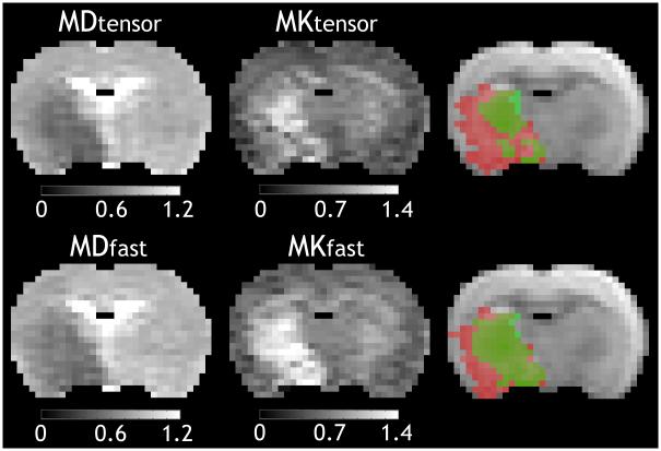
Fig. 3.
Comparison of mean diffusivity and mean kurtosis maps obtained using the conventional and the fast DKI methods. Lesions of mean diffusivity and mean kurtosis were overlaid on a diffusion-weighted image and were colored in red and green, respectively. Note that regions with concurrent diffusivity and mean kurtosis abnormalities were shown in deep green, and there were only a few pixels in the medial caudate-putamen showing kurtosis abnormality without diffusion deficit (light green). The unit of diffusivity is μm2/ms.

