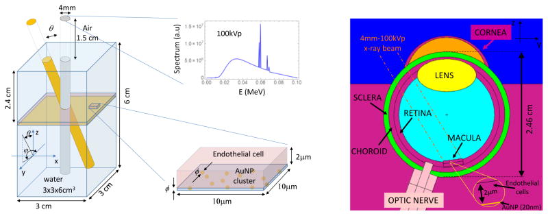
Figure 1.
(left) Simplified MC simulation geometry with the 4 mm – 100 kVp x-ray beam direction and spectrum; a zoom of the endothelial cell with at the bottom the AuNP layer is shown, (right) detailed eye geometry (YZ section), AuNP layer (20 nm) is located at the bottom of macular endothelial cells (2 μm). Neighboring organs (optic nerve, Retina, Lens, etc) and the direction of the incoming x-ray beam are depicted.

