Fig 1.
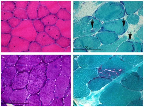
a) Cross section of gluteal muscle from a pro-ER horse (prospective exertional rhabdomyolysis) containing numerous fibres with one or more centrally displaced nuclei (haematoxylin and eosin ×40). b) Cross section of gluteal muscle from a pro-ER horse demonstrating fibres with a wavy disrupted pattern of myofibre alignment (periodic acid-Schiff (PAS) ×40). c) Cross section of semimembranosus muscle from a retro-ER (retrospective exertional rhabdomyolysis) horse showing abnormal amorphous basophilic material in several fibres (arrows) (modified Gomori trichrome ×40). d) Cross section of semimembranosus muscle from a retro-ER horse demonstrating rimmed vacuoles within a muscle fibre (modified Gomori trichrome ×40).

