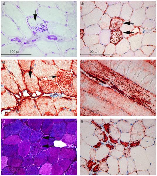
Fig 3.
a) Cross section of semimembranosus muscle from a retrospective exertional rhabdomyolysis (retro-ER) horse stained with amylase periodic acid-Schiff (PAS) demonstrating amylase-resistant abnormal PAS positive material (arrow). (PAS ×40). b) Serial frozen section demonstrating that myofibres with abnormal PAS positive material (vertical arrow) do not contain desmin positive aggregates (horizontal arrow) (desmin immunohistochemistry ×40). c) Cross section of gluteal muscle from a prospective ER (pro-ER) horse demonstrating normal staining for glycogen (PAS ×40). d) Serial formalin-fixed section of the same muscle shown in (c) showing that muscle fibres with desmin positive aggregates have normal glycogen staining (horizontal arrows in (b) and (c); desmin immunohistochemistry ×40). e) Longitudinal section of a formalin-fixed gluteal muscle from a pro-ER horse showing aggregates of cytoplasmic desmin throughout the length of a mature muscle fibre (desmin immunohistochemistry ×20). f) Cross-section of formalin-fixed gluteal muscle from a pro-ER horse showing small anguloid regenerating fibres with centrally located nuclei that have a uniform dark desmin stain (desmin immunohistochemistry ×40).

