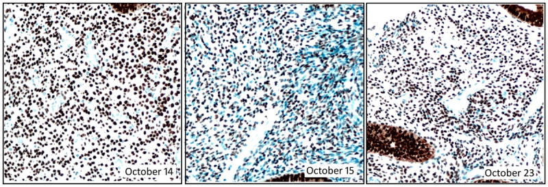
Figure 4.

Photomicrographs of the PR control (normal endometrium) from 3 consecutive days of PR staining. The dates (in 2014) are indicated with each photomicrograph. The October 15 image shows a markedly stronger counterstain, highlighting many more unstained cells than the preceding or subsequent days. Magnification 200X.
