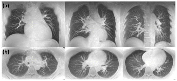
Figure 4.
MIP (25 mm slab thickness) of respiratory triggered free breathing lung images obtained at three different levels for (a) coronal and (b) axial scans. Native resolution was 1.5 × 1.5 × 5 mm3 for both scans. Scan time was 5:06 and 6:54 for complete coverage along the coronal and axial directions, respectively.

