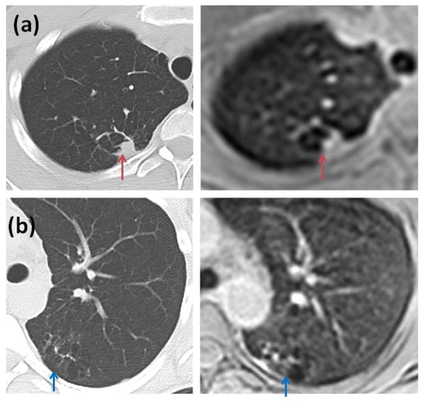
Figure 6.
Scarring or atelectasis exhibited by a patient (CT and MRI done sequentially). (a) CT image compared with lower resolution breath-hold axial MRI image (red arrows) (b) CT image compared with free breathing axial MRI image (blue arrows). Although CT offers higher resolution, MR images provide comparable visualization.

