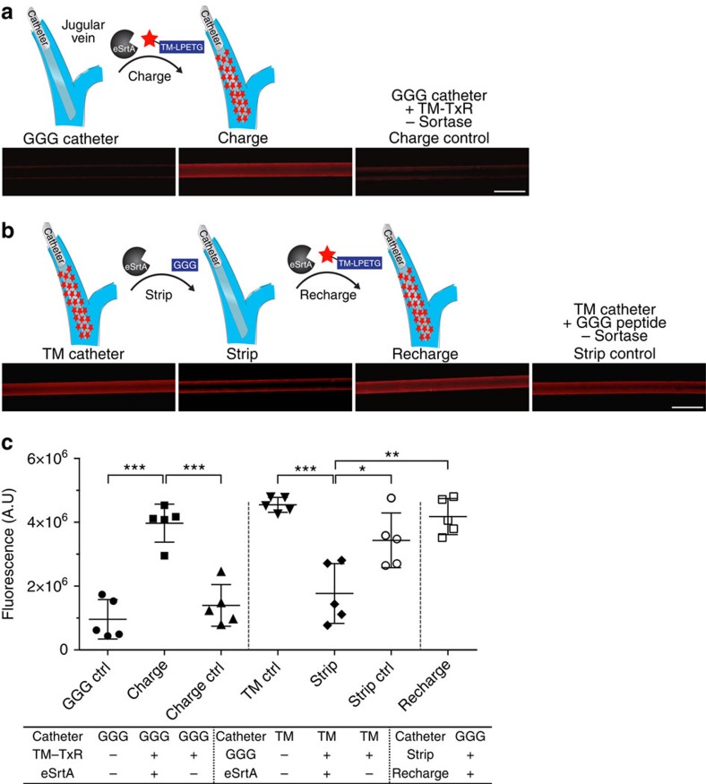
Figure 5. In situ molecular recharging of venous catheters after in vivo deployment.
(a) To characterize in situ charging of a chronically implanted venous catheter, pentaglycine-modified polyurethane catheters were initially deployed in the rat jugular vein for 7 days. Texas Red-labelled TMLPETG with or without eSrtA was systemically administered by intravenous injection via the dorsal penile vein. Catheters were removed 1 h later and imaged using fluorescence microscopy. Representative images are shown (scale bar, 1 mm). (b) To characterize in situ stripping, pentaglycine catheters linked to Texas Red-TM were deployed in the rat jugular vein for 7 days. GGG with or without eSrtA was delivered intravenously and 1 h later catheters were removed and imaged. To examine in situ recharging, Texas Red-TM catheters were first stripped by intravenous administration of GGG and eSrtA and 24 h later, Texas Red-TMLPETG and eSrtA was administered intravenously. Catheters were removed 1 h later for imaging. Representative images are shown (scale bar, 1 mm). (c) Catheter fluorescence intensity was quantified using ImageJ and plotted as. *P<0.05, **P<0.01, ***P<0.001 (Student's t-test), error bars denote s.d. (n⩾3). A.U., arbitrary units.

