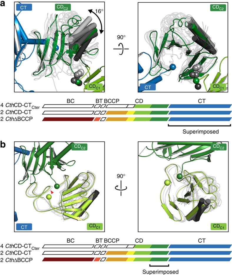
Figure 3. Variability of the connections of CDC2 to CT and CDC1 in fungal ACC.
(a) Hinge properties of the CDC2–CT connection analysed by a CT-based superposition of eight instances of the CDC2-CT segment. For clarity, only one protomer of CthCD-CTCter1 is shown in full colour as reference. For other instances, CDC2 domains are shown in transparent tube representation with only one helix each highlighted. The range of hinge bending is indicated and the connection points between CDC2 and CT (blue) as well as between CDC1 and CDC2 (green and grey) are marked as spheres. (b) The interdomain interface of CDC1 and CDC2 exhibits only limited plasticity. Representation as in a, but the CDC1 and CDC2 are superposed based on CDC2. One protomer of CthΔBCCP is shown in colour, the CDL domains are omitted for clarity and the position of the phosphorylated serine based on SceCD is indicated with a red triangle. The connection points from CDC1 to CDC2 and to CDL are represented by green spheres.

