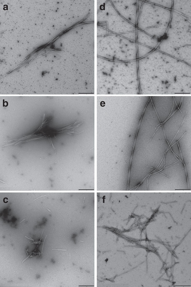
Figure 2. Effect of high pressure on Aβ fibrils.
Electron micrographs of npAβ (a–c) and pS8Aβ (d–f) aggregates, before (a,d) and after (b–c,e–f) application of high hydrostatic pressure (2,000 bar, 18 h). Many fibrils were unaffected by the high pressure (b,e), but some broken fibrils were also observed (c,f). Scale bar, 200 nm.

