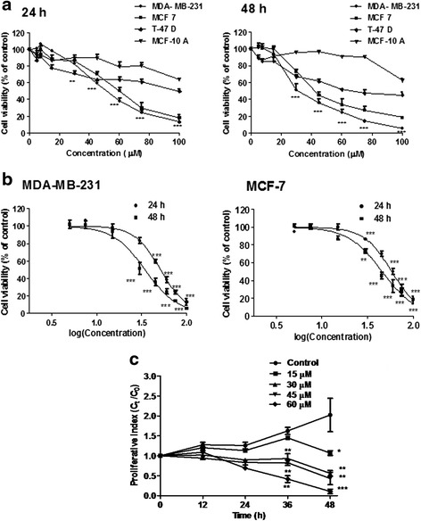
Fig. 1.

Assessment of cell viability in response to andrographolide. a Three breast cancer cell lines, MDA-MB-231, MCF-7, T-47D and a normal breast epithelial cell line MCF-10A were treated with andrographolide (5–100 μM) or vehicle control (0.1 % DMSO) for 24 h and 48 h. Time- and concentration- dependent inhibitory effects of andrographolide were evaluated by MTT assay. b The IC50 of andrographolide on MDA-MB-231 and MCF-7 were calculated by plotting the percentage of viable cells against log (concentration) of andrographolide. c Effect of andrographolide on proliferation kinetics of MDA-MB-231 cells. Cells (1 × 10⁴/well) in a 24-well culture plate were treated with varying concentrations of andrographolide for different time points. Cell proliferation was evaluated using trypan blue. Values are mean ± S.D. of three independent experiments (*P < 0.05, **P < 0.01 and ***P < 0.001)
