Fig. 2.

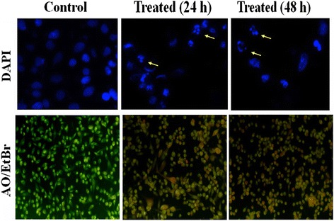
Assessment of cell morphology of MDA-MB-231 treated without or with andrographolide. Upper panel Effect of andrographolide in MDA-MB-231 cells with condensation and fragmentation of the nuclei identified by DAPI by Fluorescence microscopy. Cells were treated with IC50 concentration of andrographolide for 24 and 48 h and stained with DAPI. The fragmented apoptotic nuclei were shown by arrow. Data shown are from a representative of triplicate experiments. Lower panel Acridine orange/Ethidium bromide (AO/EtBr) staining of MDA-MB-231 cells after treatment of IC50 concentration of andrographolide for 24 and 48 h and compared with untreated control cells using fluorescence microscopy. Cells showing bright orange fluorescence indicate apoptosis in comparison to control cells showing green fluorescence
