
Fig. 8.
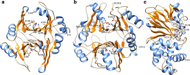
Three dimensional homology model structure of soybean glyoxalase proteins. Structures of GmGLYI-3 (a), GmGLYI-16 (b) and GmGLYII-5 (c) were built using Swiss-model server based on available close similar structure from Protein Data Bank (PDB) Zea mays GLYI (5D7Z), mouse GLYI (4OPN), and AtGLYII-2 (2Q42) proteins, respectively. All the α-helices were marked with orange colour, while β-sheets were marked with cornflower blue. The active sites residues were identified based on the alignment with template structure and shown by ball-stick model. The structure and active residues were visualized and generated using chimera program