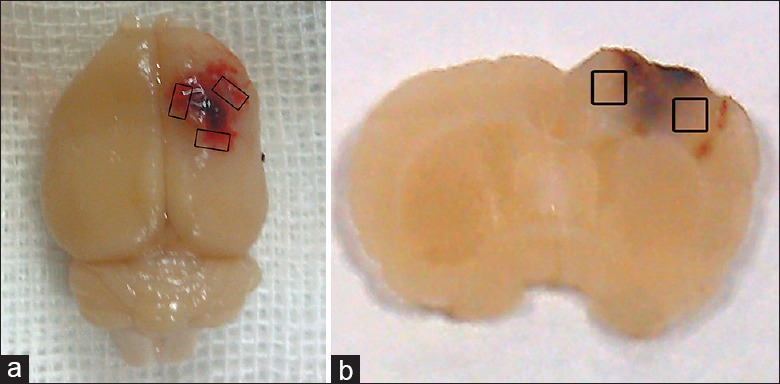
Figure 1.

Schematic representation of the sample area for detection. (a) Image of a representative TBI brain showing cortex contusion and the pericontusional region surrounding the injured cortex. The sample area for detection is marked with the black boxes. TBI: Traumatic brain injury; (b) a coronal section of a rat brain used for immunofluorescent staining and TUNEL analysis. The black boxes indicate the detected region. TUNEL: Terminal deoxynucleotidyl transferase-mediated dUTP nick-end labeling.
