Figure 3.

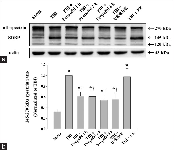
Western blot analysis of αII-spectrin in the pericontusional cortex at 24 h following moderate TBI. (a) Brain tissue lysates were immunoblotted with anti-αII-spectrin (top) and anti-β-actin (bottom) antibodies, respectively. The lanes were loaded with protein from the Sham group (Sham), the TBI group (TBI), the propofol 1 h group (TBI + Propofol 1 h), the propofol 2 h group (TBI + Propofol 2 h), the propofol 4 h group (TBI + Propofol 4 h), the lipid peroxidation inhibitor U83836E group (TBI + U83836E) and the vehicle control fat emulsion group (TBI + FE). TBI: Traumatic brain injury; (b) quantitative analysis for the ratio of the calpain-mediated 145-kDa spectrin breakdown product (SBDP) to intact 270-kDa αII-spectrin. The densitometric ratio was normalized against the TBI group. The results were expressed as the means ± standard deviation (SD) (n = 6). *P < 0.01 vs. Sham group; †P < 0.01 vs. TBI group.
