
Figure 3.
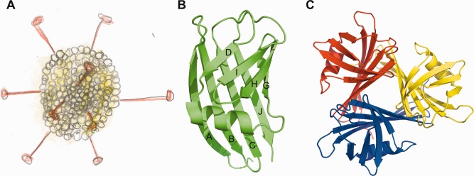
Snake Adenovirus 1 and its fiber head protein. (A) Schematic drawing of an icosahedral adenovirus with trimeric fiber proteins protruding from each of the twelve vertices. The head domains are located at the distal ends of the fibers. (B and C) Cartoon representation of a fiber head monomer (A) and a fiber head trimer (C). In part B the β‐strands are labeled. Parts B and C were prepared using the PyMOL Molecular Graphics System, Version 1.4.1, Schrödinger LLC and were first published in Singh 2014.17