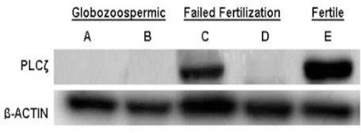
Figure 6.

Western blot analysis for PLCζ compared with β-ACTIN. Upper panel shows PLCζ western blotting of five semen samples from two globozoospermic (A and B), two non-globozoospermic patients (C,D) and one fertile individual (E) respectively. Lower panel is B-actin staining of the same individual to confirm integrity of protein content related to immunoblotting of the respective samples with an antibody against β-ACTIN. For simplicity of presentation PLCζ the extra non-specific band were crop out and only 70kDa was presented
