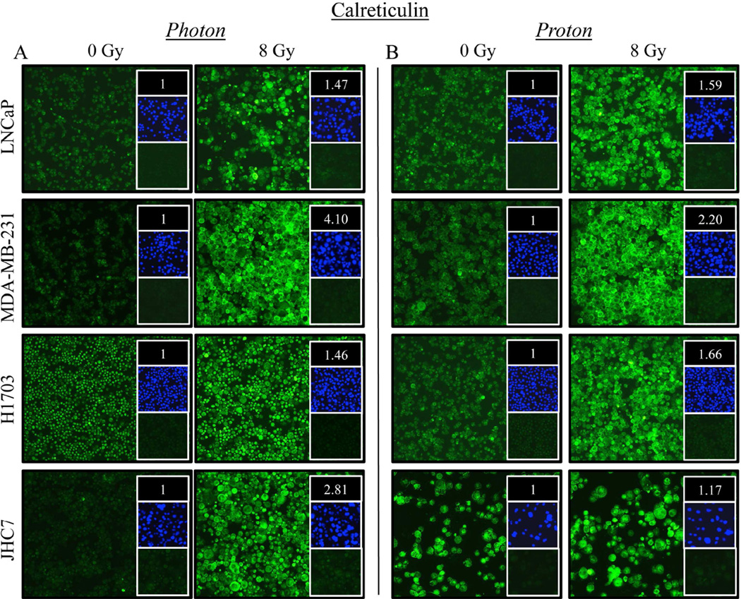
Figure 3. Carcinoma cells recovering from exposure to photon or proton radiation have increased surface and intracellular expression of calreticulin.
Calreticulin (green) expression was examined by immunofluorescence (10× magnification) 96 h after a single 8-Gy dose of (A) photon or (B) proton radiation of human prostate (LNCaP), breast (MDA-MB-231), lung (H1703), or chordoma (JHC7) cells. Upper inset: MFI normalized to that of mock-irradiated controls. Middle inset: DAPI nuclear stain (blue). Lower inset: isotype control. Data are representative of 2 independent experiments. This experiment was repeated 2 times with similar results.

