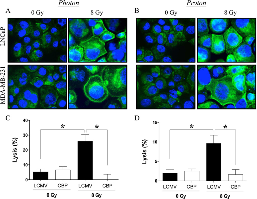
Figure 4. Carcinoma cells recovering from exposure to photon or proton radiation show increased calreticulin expression on the cell surface, resulting in heightened sensitivity to CTL-mediated killing.
Cell-surface expression of calreticulin (green) was examined by confocal immunofluorescence (10× magnification) in human prostate (LNCaP) and breast (MDA-MB-231) cells 96 h after a single dose of 8 Gy (A) photon or (B) proton radiation. Blue denotes DAPI nuclear stain. (C–D) Functional role of cell-surface calreticulin on CTL-mediated lysis. MDA-MB-231 cells were mock irradiated (0 Gy) or exposed to a single dose of 8 Gy (C) photon or (D) proton radiation. After 96 h, cells were used as targets in a CEA-specific CTL-lysis assay in the presence of calreticulin-blocking peptide (CBP, open bars) or LCMV control peptide (closed bars). Results are presented as mean ± S.E.M. from 6 replicate wells. *, statistical significance relative to controls.

