Figure 1.

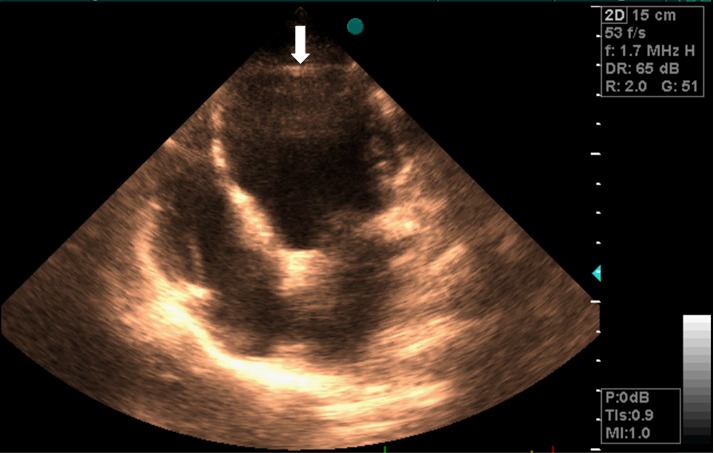
Four-chamber view in a transthoracic echocardiogram showing enlargement of the left ventricle (LV) (The LV apical structure was unclear in this view)

Four-chamber view in a transthoracic echocardiogram showing enlargement of the left ventricle (LV) (The LV apical structure was unclear in this view)