Figure 3.

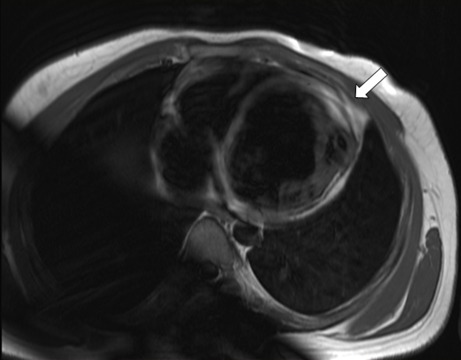
A T1-weighted image shows bright tissue replaced in the left ventricle (LV) apical position (white arrow), which could suggest the presence of fat replacement

A T1-weighted image shows bright tissue replaced in the left ventricle (LV) apical position (white arrow), which could suggest the presence of fat replacement