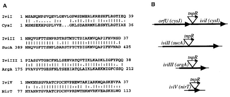
Fig. 2.

A. Similarities of deduced V. cholerae polypeptide partial sequences. Vertical lines indicate identical residues, highly conserved substitutions are represented by colons and conserved substitutions are represented by dots. Gaps were introduced into the sequences to maximize the similarities (see the Experimental procedures). For each V. cholerae polypeptide sequence shown, the C-terminal amino acid delineates the corresponding DNA site of fusion to tnpR–lacZY.
B. Schematic diagram (not to scale) of predicted V. cholerae genes containing fusions to tnpR–lacZY (designated by triangles). Transcriptional orientations are shown by the arrows.
