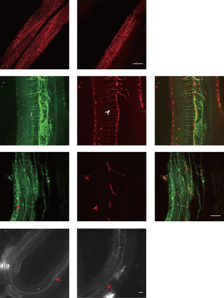
Fig. 2. Reduction of PVD 4° branches in clr-1 mutant is not caused by SAX-7/DMA-1 pathway or muscle and hypodermal integrity defects.
A-B) Confocal images of young adult animals expressing UNC-97::mCherry in muscles in wild type and clr-1(e1745) mutant, respectively. C-H) Confocal images of young adult animals expressing Pdpy-7::SAX-7s::GFP in hypodermis and PVD::mCherry in wild type and clr-1(e1745ts) mutant, respectively. The white arrow marks the SAX-7 stripes and the white arrowhead points to the quaternary dendrites. The red arrow marks the SAX-7 stripes and the red arrowhead points to the same region where no PVD 4° branches form. I-J) Confocal images of young adult animals expressing AJM-1::GFP in wild type and clr-1(e1745ts) mutant, respectively. Red arrows show the fused seam cell boundary with hypodermal cell. Scale bar is 10μm.

