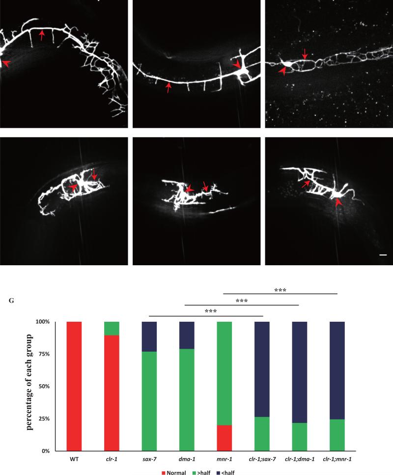
Fig. 5. clr-1 functions redundantly with the SAX-7/MNR-1 pathway to control PVD 1° branch extension.
A-F) Confocal images of young adult mutant animals expressing ser-2Prom3::GFP. Red arrows indicate the primary dendrites and red arrowheads point to cell bodies. Scale bar is 10μm. G) Quantification of primary dendrite length in wild type and different mutant backgrounds. “> half” means the length is greater than a half of a normal length of a PVD primary dendrite. “< half” means the length is less than a half of a normal length of a PVD primary dendrite. ***p < 0.001 by Chi-square test for multiple samples. n>30 for each genotype.

