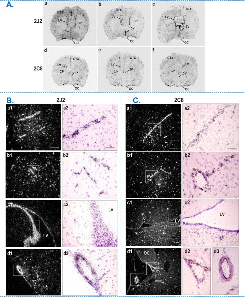
Figure 1.
Expression and distribution of human CYP2J2 and CYP2C8 mRNA in the brain of transgenic female mice by in situ hybridization (ISH). A. Bright-field film images illustrating the distribution of CYP2J2 (top) and CYP2C8 (bottom) mRNA (dark stain) from rostral to caudal in mouse forebrain transfected the respective human transcripts (a-c; d-f). B & C. Emulsion autoradiograms of human CYP2J2 (B) and CYP2C8 (C) mRNA in Tie2-CYP2J2 and Tie2-CYP2C8 mouse forebrain respectively. Photomicrographs of dark-field (a1-d1) and bright-field (a2-d2) at low and high power views, illustrating respective mRNA signal (white grains in a1-d1, dark grains in a2-d2) within coronal sections through the rostrodorsal cortex (CTX; a, b), lateral ventricular area (LV; c) and olfactory cortex in Tie2-CYP2J2 or supraoptic nucleus area in Tie2-CYP2C8 brains (OC; d). Boxed areas from a1-d1 are shown as high-power bright-field images in a2-d2, illustrating autoradiographic grains (mRNA) over blood vessels (a,b), ependymal lining of the lateral ventricle (c) and along the ventral surface of the brain (d). Scale bar = 200 μm (a1-d1) and 50 μm (a2-d2). CP, caudate putamen; CTX, cortex; LV, lateral ventricle; OC, optic chiasm; 3V, third ventricle.

