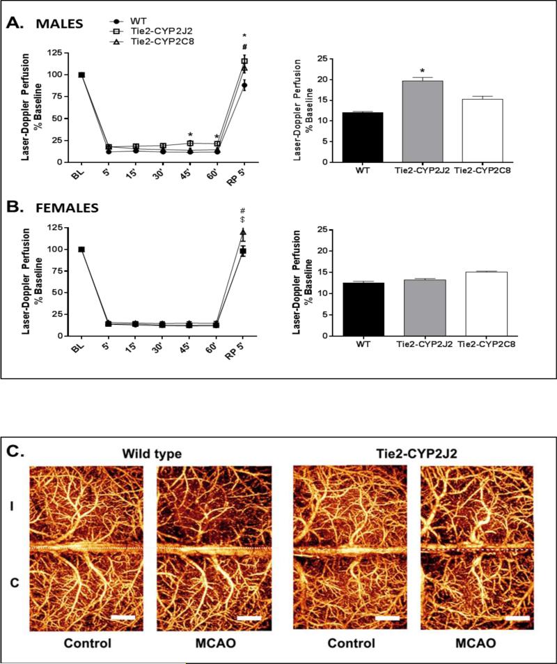
Figure 3.
Laser-Doppler cerebrocortical perfusion (LDP) during MCAO in WT, Tie2-CYP2J2 and Tie2-CYP2C8 male and female mice. A. Tie2-CYP2J2, but not Tie-2-CYP2C8 males, displayed significantly higher perfusion of the MCA territory during occlusion compared to corresponding WT males. Both Tie2-CYP2J2 and Tie2-CYP2C8 displayed higher LDP compared to WT following 5 minutes of reperfusion. B. No differences were observed in perfusion between female transgenic and WT mice during occlusion (n=10 per group). After 5 minutes of reperfusion Tie2CYP2C8 mice displayed higher LDP compared to both WT and Tie2-CYP2J2 mice. Line graph displays perfusion at 15 minute intervals during occlusion and immediately following reperfusion (RP) relative to baseline (BL). Bar graph displays average perfusion throughout the occlusion period. * and # denote p<0.05 CYP2J2 and CYP2C8, respectively, versus WT; $ denotes p<0.05 Tie2-CYP2J2 versus Tie2-CYP2C8 (n=10 per group). C. Optical microangiography (OMAG) images illustrating increased microvascular cortical perfusion of ipsilateral ischemic (I) hemisphere in male Tie2-CYP2J2 brain 24 hours after MCAO compared to WT.

