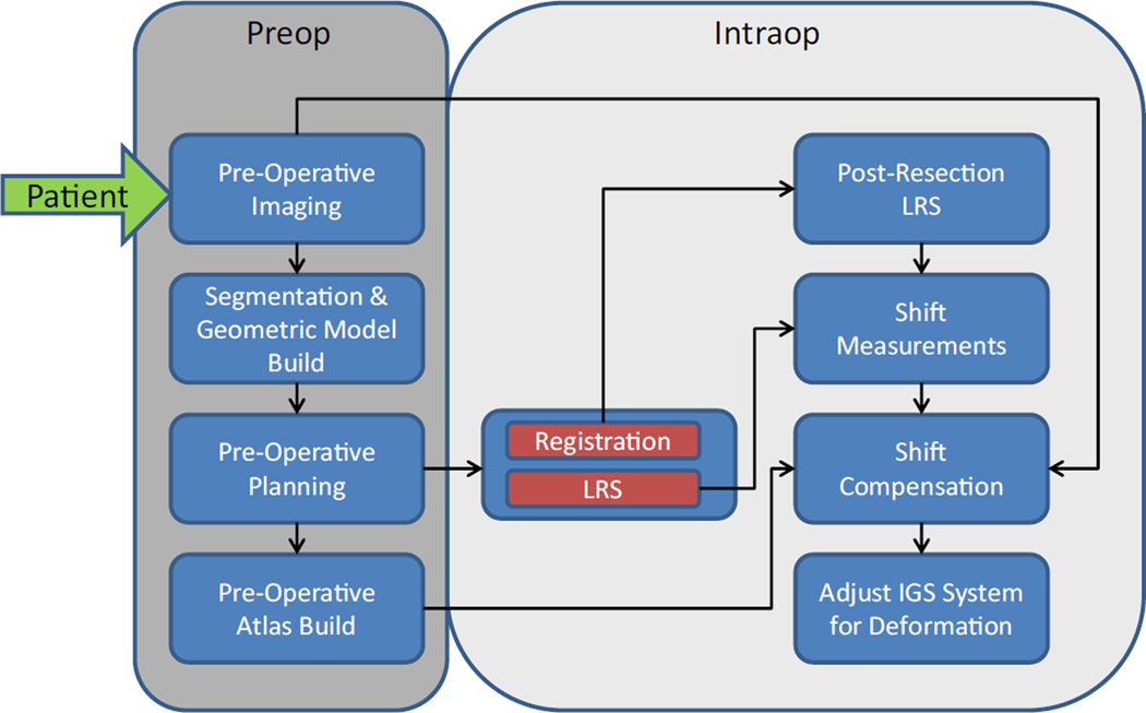
Fig. 1.
A workflow illustrating the preoperative and intraoperative computational processing steps involved in producing an updated brain shift image. The inputs are preoperative MR images, face laser range scan (LRS) for registration, and pre- and postresection cortical brain surface LRS to drive the inverse modeling

