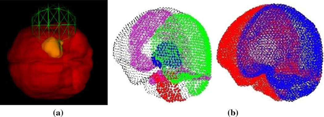
Fig. 2.
a Preoperative GUI planner with craniotomy location and size tool demonstrated, and b boundary conditions automatically generated from preoperative planning tool with (left) showing fixed brain stem nodes in red, stress-free nodes in green, cranially constrained but lateral freedom in black, with dural septa nodes of falx and tentorium shown in magenta, and tumor shown in blue on the left image. On the (right), drainage conditions are specified with drainage allowed on regions of the brain open to atmosphere in blue with nondraining nodes in red

