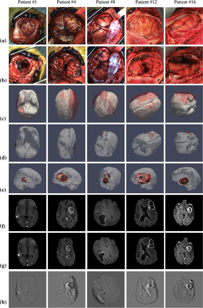
Fig. 3.
For patients #1, 4, 8, 12, and 16, illustrated are a preresection field of view bitmap (FOVBMP), b post-resection FOVBMP, c brain shift as observed by the overlay of deformed (white) and undeformed (red) brain mesh, d top view and e side view of the deformed brain mesh overlaid with the postresection LRS scans, f original MR image, g deformed MR image, and h image difference between original and deformed MR images

