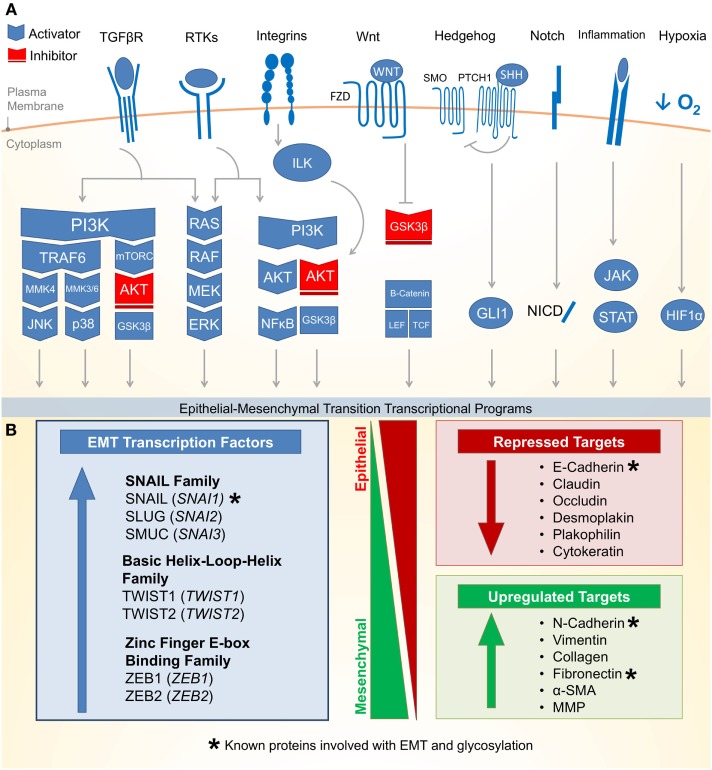
Figure 1.
Molecular pathways and targets of the epithelial–mesenchymal transition. (A) (1) One of the most well characterized EMT-inducing pathways is the transforming growth factor-β (TGF-β) family receptors capable of inducing PI3K–AKT, ERK MAPK, p38 MAPK, and JUN N-terminal kinase (JNK) pathways (activating pathways in blue; inactivating pathways in red). (2) The RAS–RAF–MEK–ERK MAPK pathway lies downstream of a number of growth factor activated receptor tyrosine kinases (RTKs) and activates a number of major EMT transcription factors (TFs). (3) Integrin signaling can have a multipronged effect on EMT by both interrupting critical epithelial adhesion molecules (e.g., E-cadherin) and antagonizing GSK3-β via the integrin-linked kinase (ILK)-AKT signaling, thus promoting EMT. (4) WNT signaling can also interfere with GSK3-β, thus stabilizing β-catenin to promote EMT transcriptional programs in cooperation with lymphoid enhancer-binding factor 1 (LEF1) and T-cell factor (TCF). (5) The Hedgehog (HH)-glioma 1 (GLI1) and (6) NOTCH pathway both can promote transcription of the EMT regulators. (7) Recently, a number of inflammatory pathways downstream of interleukin (IL) signaling (e.g., IL-6) have demonstrated the activation of the Janus-kinase (JAK)-signal transducer and activator of transcription 3 (STAT3) pathway, which in turn promotes EMT transcription factors. (8) Hypoxia is capable of activating a number of key components of EMT through the hypoxia-induced factor 1 (HIF1α). (B) Downstream of these signal transduction pathways leading to EMT are a variety of transcription factors with the ability to alter epithelial gene expression. As an epithelial plasticity program, many of the target genes altered include adhesion molecules. Known glycosylated proteins involved with EMT are denoted with an asterisk (*).

