Figure 1.
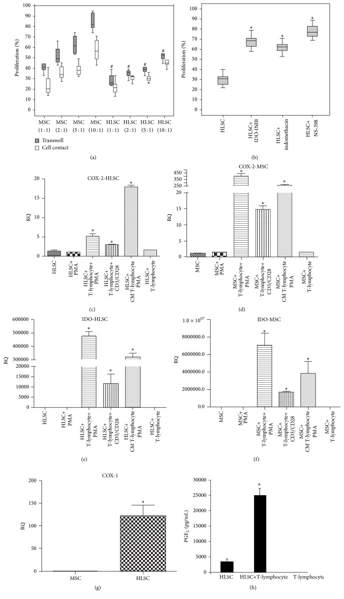
HLSCs suppress T-lymphocyte proliferation through IDO and PGE2 activity. (a) CD3+ lymphocytes were stimulated with PMA, in the presence or the absence of MSCs or HLSCs at different ratio (lymphocytes: HLSCs/MSCs 1 : 1, 2 : 1, 5 : 1, and 10 : 1) in direct contact or in the presence of transwells. The proliferation rate was evaluated after 3 days of coculture. Results are expressed as mean ± SD of 6 different experiments conducted in duplicate. 100% of proliferation corresponds to lymphocytes stimulated with PMA. Data were analysed by nonparametric Wilcoxon test: ∗ p < 0.05 lymphocytes stimulated with PMA cocultured in contact with HLSCs versus lymphocytes stimulated with PMA cocultured in contact with MSCs; # p < 0.05 lymphocytes stimulated with PMA cocultured with HLSCs, in the presence of transwells, versus lymphocytes stimulated with PMA cocultured with MSCs, in the presence of transwells. (b) IDO and PGE2 are involved in HLSC suppression of T-cell proliferation. CD3+ cells were stimulated with PMA in the presence of HLSCs (5 : 1 ratio) with or without inhibitors of IDO (1-methyl-L-tryptophan) and Cox-1 and/or Cox-2 (indomethacin, NS-398) in the presence of transwells. The proliferation rate was evaluated after 3 days. Results are expressed as mean ± SD of 6 different experiments in duplicate. 100% of proliferation corresponds to lymphocytes stimulated with PMA. Data were analysed by ANOVA with Bonferroni correction: ∗ p < 0.05 lymphocytes stimulated with PMA cocultured with HLSCs and different inhibitors versus lymphocytes stimulated with PMA cocultured with HLSCs. (c) and (d) mRNA expression levels of Cox-2 were evaluated in HLSCs (c) and MSCs (d) unstimulated, stimulated with PMA, after 3 days of coculture with CD3+ lymphocytes stimulated with PMA or with CD3/CD28 antibodies, stimulated with CM from T-cells stimulated with PMA, and cultured with unstimulated lymphocytes. (e) and (f) mRNA expression levels of IDO were evaluated in HLSCs (e) and MSCs (f) unstimulated, stimulated with PMA, after 3 days of coculture with CD3+ lymphocytes stimulated with PMA or with CD3/CD28 antibodies, stimulated with CM from T-cells stimulated with PMA, and cultured with unstimulated lymphocytes. Cox-2 and IDO expression levels increased after coculture with activated lymphocytes and after stimulation with CM. Data are expressed as relative quantification using ΔΔCt method. Normalization was made using actin as housekeeping gene. Data were analysed by ANOVA with Bonferroni correction, ∗ p < 0.05 gene expression of MSCs or HLSCs after coculture with CD3+ cells stimulated with PMA or CD3/CD28 antibodies or after culture with CM versus MSCs or HLSCs alone. (g) mRNA expression levels of COX-1 were evaluated in MSCs and HLSCs. Data are expressed as relative quantification using ΔΔCt method. Normalization was made using actin as housekeeping gene. Data were analysed by Student's t-test (unpaired, 2-tailed); ∗ p < 0.05 COX-1 expression of HLSCs versus MSCs. (h) Cell supernatants from cocultures were harvested to detect PGE2 production by ELISA. A significant increased production of PGE2 (p < 0.05) was observed after 3 days of coculture of CD3+ cells stimulated with PMA and cocultured with HLSCs.

