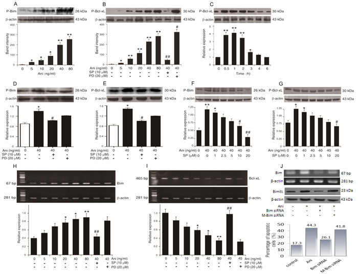
Figure 3. Anisomycin promoted the apoptosis of Jurkat T cells through the JNK-dependent activation of Bim/Bcl-xL.
Jurkat T cells were pre-incubated with 10 μM SP600125 (SP), 20 μM PD98059 (PD) or the increasing concentrations of SP600125 for 2 h before treated with the indicated concentrations of anisomycin (Ani) for 6 h. Additionally, the cells were treated with 40 ng/ml of anisomycin at the indicated time points. (A–G) The expression levels of P-Bim and P-Bcl-xL were assessed by Western blot analysis. (H,I) The alterations of Bim and Bcl-xL mRNAs were evaluated by RT-PCR. (J) Furthermore, Bim-targeting siRNA was used to knock down the bim gene to confirm the correlation of its mRNA and protein levels with the cell apoptosis. Data are presented as the mean ± SD of three independent experiments for (A–I). *P < 0.05, ** P < 0.01 vs. the untreated control, #P < 0.05, ##P < 0.01 vs. the 40 ng/ml of anisomycin alone.

