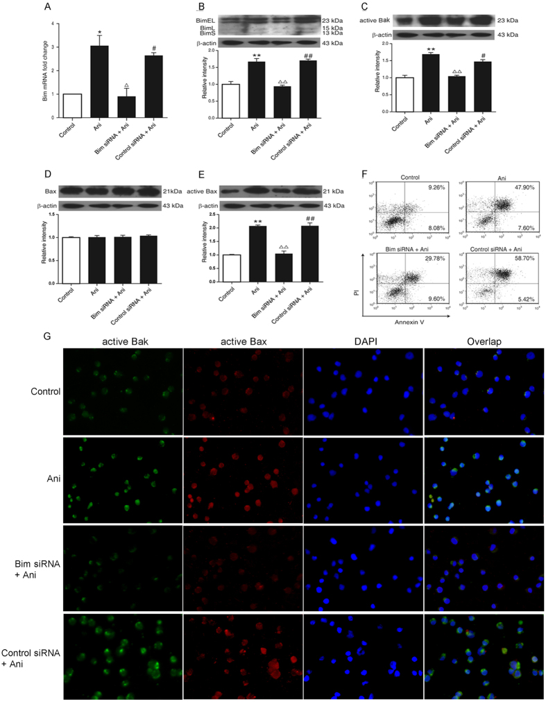
Figure 7. Role of Bim in the anisomycin-induced apoptosis in Jurkat T cells.
The Jurkat T cells were transfected with 100 nM of Bim-targeting siRNA or control siRNA for 24 h to knockdown the bim gene. Then, 40 ng/ml of anisomycin was added into the cells for 24 or 48 h. (A) The Bim mRNA expression was evaluated by the real-time qRT-PCR. (B) The level of Bim protein was determined by Western blotting. (C–E) The levels of active Bak, Bax and active Bax were also measured through Western blotting. (F) The apoptotic proportion of the treated cells was analyzed by flow cytometry. (G) In situ immunofluorescence staining was performed for the changes of the active Bak and Bax in the treated cells (×200). The data are presented as the mean ± SD of three independent experiments. *p < 0.05 and **p < 0.01 vs. the untreated control, Δp < 0.05 and ΔΔp < 0.01 vs. the anisomycin group, #p < 0.05 and ##p < 0.01 vs. the Bim siRNA group.

