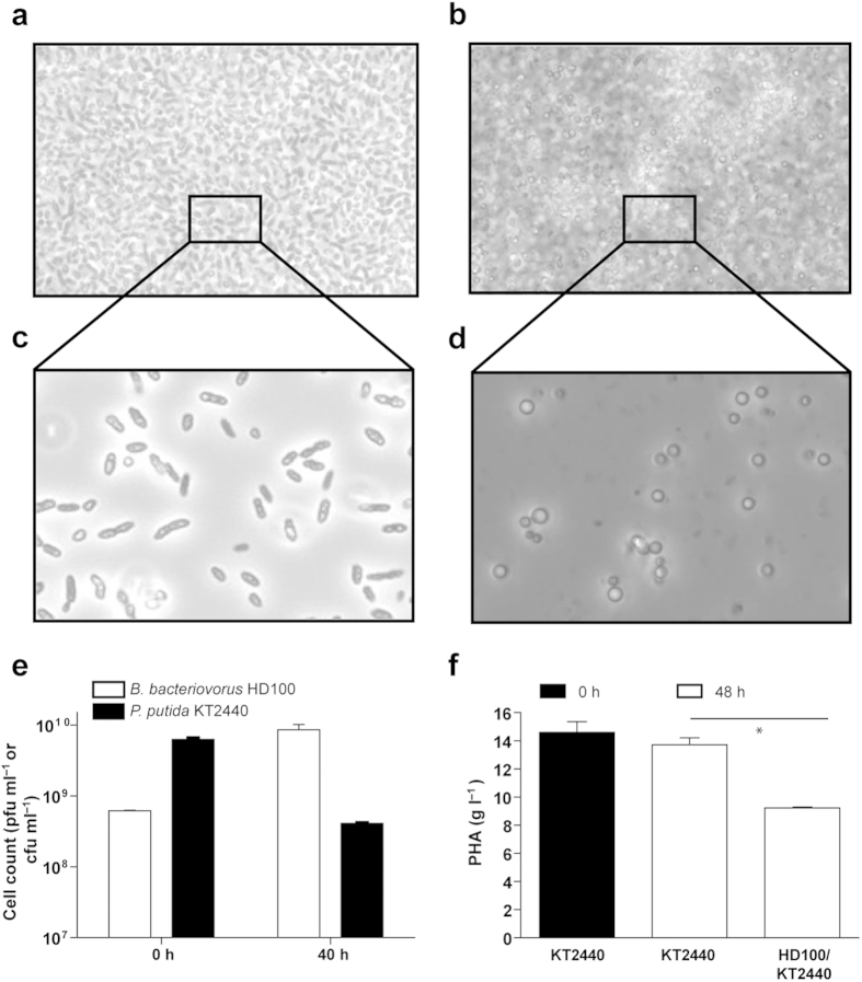
Figure 3. B. bacteriovorus HD100 preying on high cell densities of P. putida KT2440 accumulating mcl-PHA.
(a) Phase-contrast microscopy of a co-culture of B. bacteriovorus HD100 preying on P. putida KT2440 at the onset of predation (time zero) and (b) after 40 h of incubation. (c,d) 1:100 dilution of the co-cultures from panels (a,b), respectively. Mcl-PHA granules can be observed in the extracellular medium after 40 h of predation. (e) Cell viability assay of the co-culture of B. bacteriovorus HD100/P. putida KT2440 (white bars and black bars, respectively) at the onset of predation (time zero) and within 40 h. (f) Total PHA content in the co-culture of B. bacteriovorus HD100/P. putida KT2440 compared to the control culture (KT2440 without the predator) at the onset of predation (time zero) (white bars) and within 40 h (black bars). Asterisks (*) indicate significant differences (*P < 0.05).

