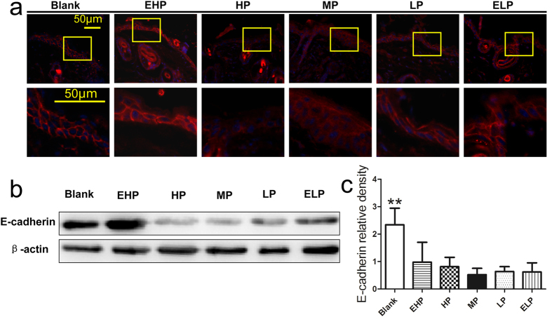
Figure 9. Migration of keratinocytes in the wound.
(a) The expression of E-cadherin at the wound edge at 3 days post-wounding, which revealed that the staining of E-cadherin at the wound edge decreased without a typical linear pattern when MP-PU membrane was applied. (b) E-cadherin and β-actin protein levels were determined by Western blot, and (c) relative densities of E-cadherin protein level in each group are shown. The values were calculated as the mean ± SD (n = 3), **p < 0.01.

