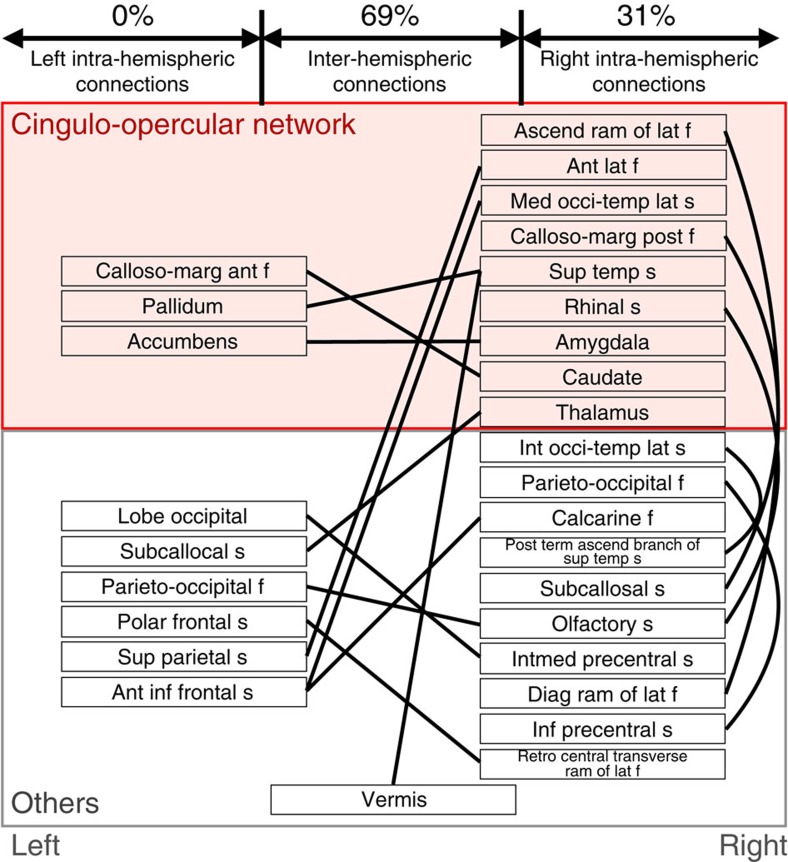
Figure 3. The 16 FCs (solid lines) and their terminal regions (names in boxes).
The left and right halves of the figure correspond to the left and right brain hemispheres, respectively. The FCs were classified into three hemispherical categories: left intra-hemispheric, right intra-hemispheric and inter-hemispheric. The terminal regions defined by the Brainvisa Sulci Atlas belong to either cingulo-opercular or other networks. The red background indicates the cingulo-opercular network. ant, anterior; ascend, ascending; calloso-marg, calloso-marginal; diag, diagonal; f, fissure; inf, inferior; int, internal; intmed, intermediate; lat, lateral; med, median; occi-temp, occipito-temporal; post, posterior; ram, ramus; s, sulcus; sup, superior; temp, temporal; term, terminal.

