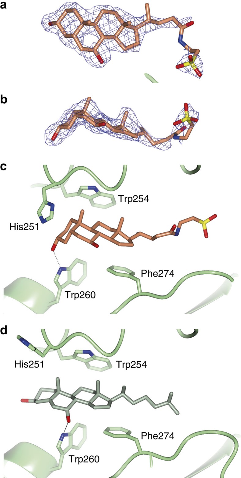
Figure 4. The bile salt TUDCA binds the tunnel of ATX.
(a,b) The 2mFo-DFc electron density map before ligand placement is contoured at 1.2 RMS and shown as a blue wireframe model in two views; TUDCA (orange carbons; oxygens in red, sulfur in yellow and nitrogen in blue) is shown as a stick model. (c,d) Comparison of the binding of TUDCA and 7α-hydroxycholesterol; Trp260 forms a hydrogen bond (dotted line) with the 3α-OH of TUDCA (in orange), whereas it forms a hydrogen bond with 7α-OH of the hydroxycholesterol (in green). In the TUDCA-bound structure, His251 flips towards the A-ring, whereas in both structures Trp260 and Phe274 pack against the steroid ring system.

