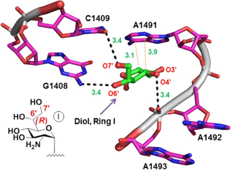
Figure 2.

Three-dimensional structure of 80S ribosome from S. cerevisiae in complex with G418 (PDB code 4U4O),8 highlighting the interactions of G418 ring I (green) into the decoding-site rRNA (magenta, E. coli numbering). The ring I oxygen atoms (red) are specified by numbers, including the modeled O7′-oxygen (PyMol software). H-bonds (dashed black lines) and C–H···π stacking (dotted red lines) are shown. The inset shows chemical structure of ring I of G418 with the modeled additional 7′-OH.
