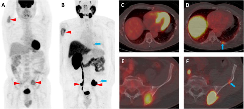
Figure 1.

A 58-year-old male patient with known clear cell RCC metastases to the skeleton was imaged contemporaneously with two PET/CT examinations, one with 18F-FDG and a second with 18F-DCFPyL, a small molecule inhibitor of PSMA. The maximum intensity projection images from the two examinations (18F-FDG, A, and 18F-DCFPyL, B) demonstrate concordance of multiple radiotracer-avid lesions including the proximal right humerus and both iliac bones (red arrowheads). However, additional subtle sites of 18F-DCFPyL uptake are noted that do not have corresponding 18F-FDG uptake (blue arrows). These sites include subtle endosteal scalloping of the left posterior ninth rib and the left iliac bone without accompanying 18F-FDG uptake (C and E, blue arrows). In contrast, the axial 18F-DCFPyL PET/CT images demonstrated moderate radiotracer uptake at these sites (D, SUVmax (lean body mass corrected) 3.2, blue arrow and F, 18F-DCFPyL SUVmax 2.7, blue arrow).
