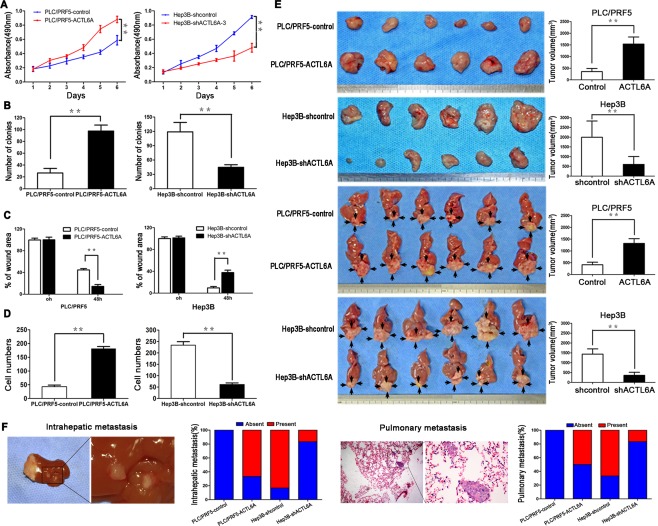
Figure 2.

ACTL6A promotes proliferation and metastasis of HCC in vitro and in vivo. (A) Proliferation of PLC/PRF5‐ACTL6A, Hep3B‐shACTL6A‐3 cells and control cells was examined by MTT and colony formation assays (B). (C) Wound‐healing assay and (D) transwell invasion assay were subjected to detect the migration and invasion capacity of ACTL6A‐interfered cells. (E) SC tumors from PLC/PRF5‐ACTL6A and Hep3B‐shACTL6A cells and their control cells are shown in the upper two panels. Orthotopic tumors from each indicated groups are shown in the lower two panels (black arrows indicate tumors). Tumor volumes of tumors are shown in the right panels. (F) Representative pictures of intrahepatic and lung metastasis; metastatic nodules proportion of livers or lungs was calculated and compared.* P < 0.05; ** P < 0.01 based on the Student t test. Error bars, standard deviation.
