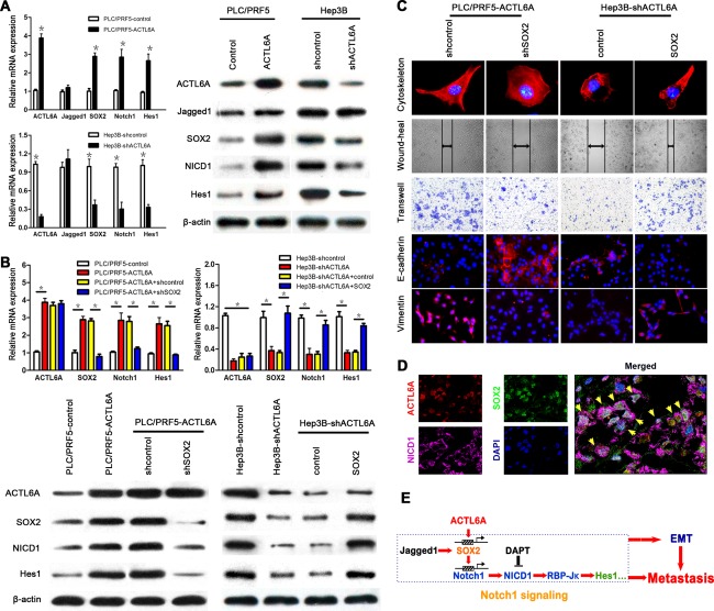
Figure 6.

ACTL6A activates Notch signaling by SOX2. (A) mRNA and protein expressions of ACTL6A, SOX2, and key members of Notch1 signaling were detected in PLC/PRF5‐ACTL6A, Hep3B‐shACTL6A, and their control cells. (B) mRNA and protein expressions of ACTL6A, SOX2, Notch1, and Hes1 in ACTL6A‐interfered HCC cells with SOX2 knockdown or ectopic expression. (C) The cytoskeleton, migration, invasion, and EMT marker expression assays of ACTL6A‐interfered HCC cells with SOX2 knockdown or ectopic expression. (D) Representative triple IF images showed that ACTL6A, SOX2, and NICD1 were colocalized in HCC tissue detected by confocal laser scanning microscope (yellow arrows indicate colocalization cells). (E) The schematic diagram of ACTL6A activating Notch1 signaling by SOX2, and promoting metastasis and EMT of HCC. Abbreviation: DAPI, 4′,6‐diamidino‐2‐phenylindole.
