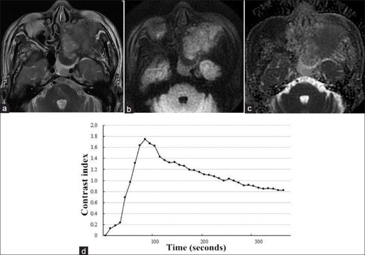
Figure 4.

(a) Axial T2-weighted magnetic resonance image in a 76-year-old man showed a left-sided tumor mass in the maxillary and ethmoid sinus with heterogeneously intermediate signal intensity. (b) On axial diffusion-weighted imaging at b = 1000 s/mm2, the mass showed limited signal loss. (c) Corresponding apparent diffusion coefficient (ADC) map showed the mass with whole slice ADCb0,1000 = 1.170 × 10⁻³ mm2/s. (d) Time-intensity curve in this patient was characterized as a washout-shaped pattern.
