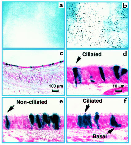
Figure 3.
Gene transfer to rabbit tracheal epithelia in vivo using FIV-βgal vector. Panels show results 5 days after gene transfer. Low magnification en face view of X-gal–stained trachea from control (a) or FIV vector–treated trachea (b). Blue cells were only seen in the trachea transduced with the FIV vector (b). (c) Low-magnification view of X-gal–stained tracheal section. β-galactosidase–expressing cells are noted at both the surface and basal cell levels of the transduced epithelium. (d–f) Higher-magnification views of tracheal epithelium showing cell types expressing β-galactosidase. No inflammatory cells were noted in control or transduced specimens (n = 4 animals). Scale bar in d also applies to e and f.

