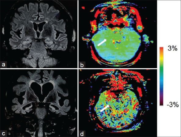
Figure 2.

Fluid attenuated inversion recovery (FLAIR) image (a) and amide proton transfer (APT)-weighted image (b) of a typical normal control (female, 72 years old, mini-mental state examination [MMSE] score 29). FLAIR image (c) and APT image (d) of an Alzheimer's disease (AD) patient (female, 70 years old, MMSE score 16). Atrophy of bilateral hippocampi (Hc), enlarged lateral ventricles and widened sulci in AD were seen on the coronal FLAIR image. The APT-weighted intensities in regions of the Hc (white arrow) were higher in AD patients than in normal controls.
