Figure 3.

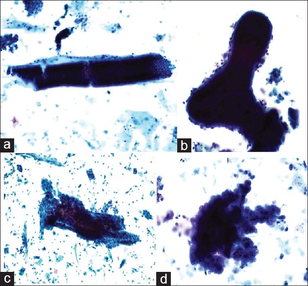
(a) Negative for endometrial lesions: Long, straight tube-shaped cell clumps with a small amount of stromal cells on the margin is the most common type of cell clumps in the proliferative endometrium, observed using a low-power microscope (Papanicolaou stain, ×20); (b) Benign endometrium: Dilated and branched cell clumps are always seen. The contour of the cell clumps is smooth and occasionally a few stromal cells can be observed (Papanicolaou stain, ×40); (c) Atypical endometrial cell: Double-layer or folded irregular cell clumps are observed (Papanicolaou stain, ×20); (d) Suspected endometrial carcinoma: Papillo-shaped bordered cell clumps with atypical cells can be observed (Papanicolaou stain, ×100).
