Figure 1.

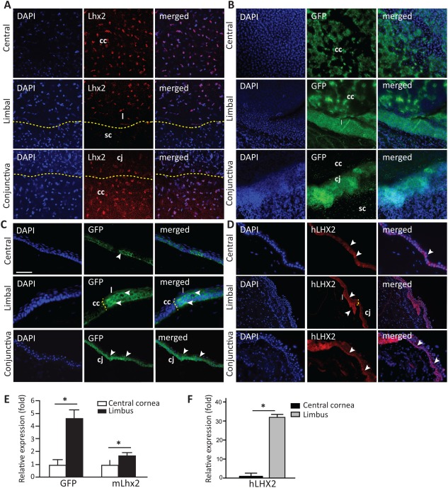
Lhx2 is expressed in the central cornea and limbus.
Lhx2 wholemount cornea and conjunctival nuclear staining (red) were detected on Lhx2eGFP corneal tissue in central, limbal, and conjunctival tissue (A). GFP expression of Lhx2eGFP in corneal wholemounts was detected in the central cornea and spanned the entire limbal region, while conjunctival tissue highly expressed Lhx2eGFP (B). In tissue cross sections, GFP‐positive cells were detected in the basal layer of the central corneal epithelium. In the peripheral cornea where the limbus is located, GFP expression was more abundant including suprabasal and basal layers. Basal cells of the conjunctiva stained positive for GFP as well (C). In human tissue, LHX2 nuclear staining in the central cornea localized to few cells in the basal layer, and included basal and suprabasal cells in the limbus. The conjunctiva distinctly expressed LHX2 in basal cells (D). Using quantitative reverse transcriptase polymerase chain reaction analysis, expression of GFP and Lhx2 was found significantly higher in murine epithelial cells originating from the limbus when compared with those from the central cornea (GFP = 4.5‐fold higher, Lhx2 = 1.6‐fold higher vs. central corneal epithelium) (E). Similarly, human LHX2 was expressed at significantly higher levels in limbal versus central corneal epithelium (32‐fold higher) (F). White arrows indicate Lhx2eGFP + and hLHX2 + cells, respectively; yellow dashed line separates the central cornea, from the limbus, conjunctiva, and sclera. Scale bar = 50 µm for images in (A–D). Data are means ± SE (human n = 3, mouse n = 4). *, p ≤ 0.05. Abbreviations: cc, central cornea; cj, conjunctiva; DAPI, * * *; GFP, green fluorescent protein; l, limbus; sc, sclera.
