Figure 3.

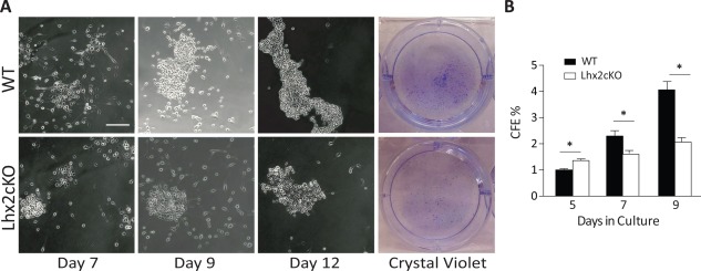
Lhx2cKO‐derived corneal epithelial cells had a reduced capacity to form colonies in vitro freshly isolated WT and Lhx2cKO corneal epithelial cells were seeded in six‐well plates and allowed to form colonies as indicated in Material and Methods section. Phase contrast and crystal violet staining showed that Lhx2cKO‐derived cells have a reduced capacity to form colonies when compared with WT cells (A). Initially Lhx2cKO‐derived cells formed more colonies at day 5 in culture, after day 9, they only reached 50% of those formed by WT‐derived cells. Quantification of the colony forming efficiency on crystal violet staining showed a significant decrease in Lhx2cKO compared with WT mice (B). Scale bar = 200 µM. Data are means ± SE (n = 3). *, p ≤ 0.05. Abbreviations: CFE, colony forming efficiency; WT, wild type.
