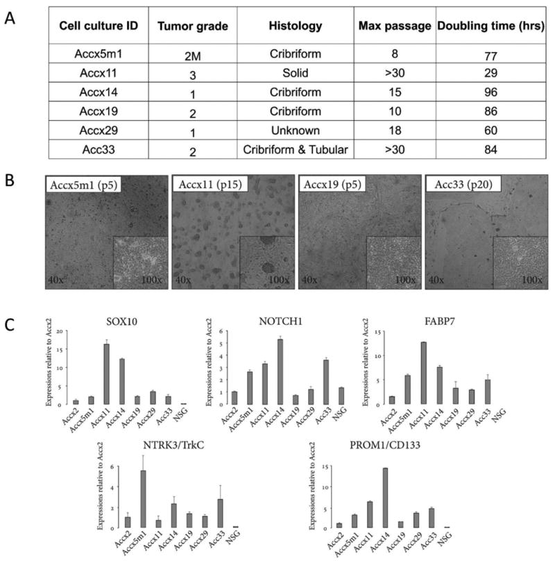
Figure 1. Clinical, cytological, and molecular properties of ACC cell cultures.

(A) ACC cell cultures produced from PDXs (Accx) and one clinical specimen (Acc33). M, metastases. (B) Low and high magnification brightfield images of cultured ACC cells at indicated passages for Accx5m1, Accx11, Accx19, and Acc33. (C) Real-time PCR quantification (qRT-PCR) of gene expression for SOX10, NOTCH1, FABP7, NTRK3/TrkC, and PROM1/CD133 in cultured ACC cells compared to cultured cells isolated from normal salivary gland (NSG). In all qRT-PCR experiments, expression is normalized to β-actin and error bars show standard errors representative of at least two independent experiments.
