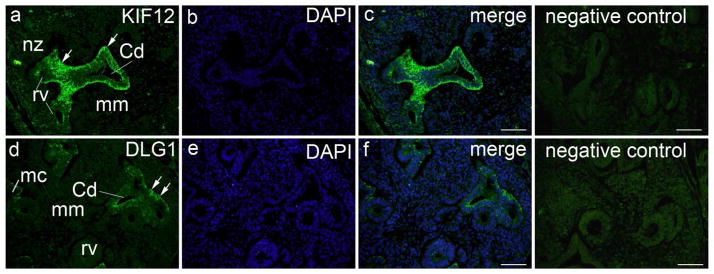
Figure 2. Expression of KIF12 and DLG1 in the developing human kidney.
Transversal section through lumbosacral part of human embryo (6th week of development): a) within the nephrogenic zone (nz), KIF12 is weakly expressed in the developing nephron (renal vesicle – rv) and negative in the metanephric mesenchyme (mm). KIF12 is strongly expressed (arrows) in the UB stalk and UB-derived structures, such as the epithelium of collecting ducts (Cd), while the surrounding mesenchyme is negative; b) DAPI nuclear staining; c) merge of a and b; negative isotype control. d) DLG1 is weakly or not expressed in the developing nephron (renal vesicle – rv, metanephric cup - mc) and negative in the metanephric mesenchyme (mm), while it is moderately expressed (arrows) in the epithelium of collecting ducts (Cd); e) DAPI nuclear staining; f) merge of d and f; negative isotype control. Immunostaining of Kif12 and Dlg1, magnification ×40, scale bar 25Xm.

