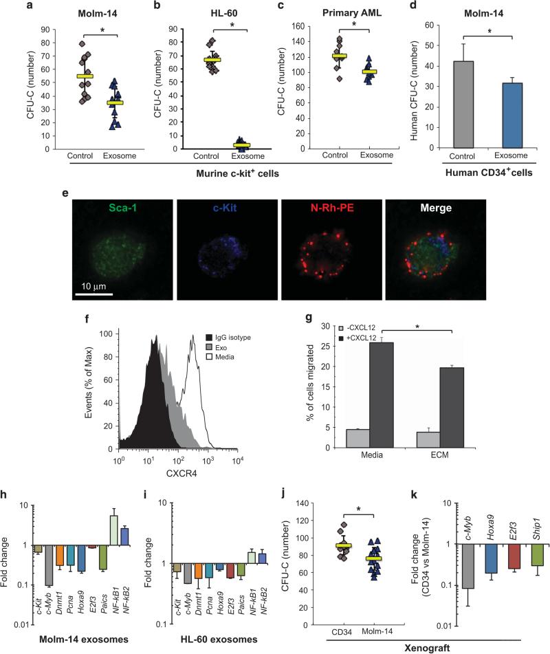
Figure 3.
AML exosomes modulate the function of HSPC. CFU-C assay for murine c-Kit+ progenitor cells after in vitro exposure to exosomes from Molm-14 (a) or HL-60 cells (b) cultured under hypoxia for 48 h. 10% VF-FBS medium was used as the control. (c) CFU-C for murine c-Kit+ progenitor cells after 48-hr in vitro exposure to exosomes from AML primary cells. (d) CFU-C for human CD34+ cells after 48 h in vitro exposure to exosomes from Molm-14. (e) Murine lin- cells were exposed to N-Rh-PE labeled Molm-14 exosomes overnight, labeled for Sca-1 and c-Kit, and imaged using deconvolution microscopy. (f) Expression of CXCR4 on lineage-depleted BM cells after 24-h exposure to Molm-14 exosomes. (g) Migration of lineage-depleted bone marrow cells along a CXCL12 gradient after 24-h culture with Molm-14 ECM or media alone. (h and i) Hematopoietic gene profile of c-Kit+ cells was examined by qRT-PCR after exposure to exosomes from Molm-14 or HL-60. (j) CFU-C for murine c-Kit+ progenitor cells collected from animals engrafted with Molm-14 (n = 6) or human CD34+ cells (n = 4). (k) Gene expression in c-Kit+ progenitor cells from xenografted animals, normalized to Gapdh. Data are representative of three independent experiments. *P < 0.01.

